Freeze frame: Scientists use new electron microscope to explore the mysteries of life

In a winding corridor behind a loading dock in the basement of Arizona State University's Schwada building, a group of ASU scientists are meeting in a lab, deeply focused, exploring the mysteries of life.
Tucked in the basement in the heart of the Tempe campus is the only microscope of its kind in Arizona, ASU's Titan Krios, a dedicated cryogenic transmission electron microscope (or cryo-EM) that uses flash-frozen samples to explore the complexities of cellular life.
Associate Research Scientist Dewight Williams is the maestro behind the operations, serving as a collaborator to all who want to solve burning biological questions. He described the project’s mission as solving “the assembly states of cellular life ... that create cells that can move and crawl and sense and do all this amazing stuff we consider life.” By revealing the structures of the true building blocks of organisms — proteins — the lab opens up new vistas of discovery.
One of ASU’s best kept secrets is kept in the basement for a reason: to keep a controlled and stable environment for the best possible microscopic imagery. The floor is made of a 4-foot-deep slab of steel-reinforced concrete in order to prevent the slightest seismic vibration that could corrupt the images. As the scientific director of the microscope, Williams noted that “to understand how biology works, you understand how the atoms are arranged ... it’s all molecules interacting with molecules. So if you can understand the structure of those molecules, probable interactions and how they do what they do ... that requires high-resolution information.”
Most of the scope functions are automated, thus allowing the scientists to multitask while the scope takes as many as 5,000 images in a given day at a resolution of 1.4 angstroms (an angstrom is the width of the smallest element, hydrogen). Scientists need only prepare samples, load the tray and occasionally realign the lenses.
The data is collected by flooding the sample with a 300,000-volt electron beam and creating a small probe that collects reflected light like a TV image composed of individual pixels. By capturing images of proteins from various angles, they can develop a 3D image of its structural makeup.
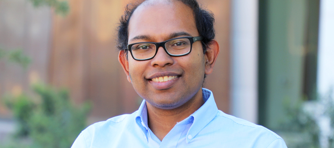
Abhishek Singaroy
Faculty from a wide range of universities and fields have requested time with the cryo-EM to help build a new picture of the structures of cellular life at work. By solving one protein structure at a time, they are building, protein brick by brick, a new picture of the structures of cellular life at work.
The cryo-EM microscope continues to yield new information and is currently part of Assistant Professor Abhishek Singharoy’s project to uncover the cellular process associated with SARS-CoV-2 infection.
His experience in molecular dynamics and kinetic modeling have propelled him to explore the prominent topic. He hopes to uncover the underlying mechanisms that make up the virus.
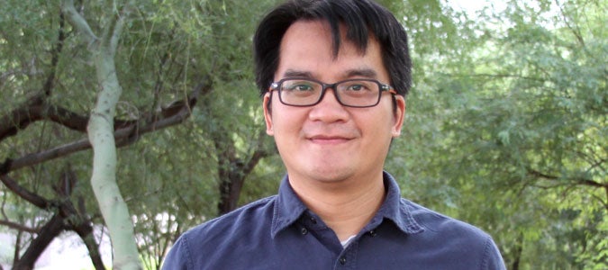
Po-Lin Chiu
ASU Assistant Professor Po-Lin Chiu is a biophysicist and frequent user of the machine. His lab utilizes the machine for examining the impact of various proteins on brain health, allowing Chiu to search for signs of neurodegeneration.
The high atomic resolution of the microscope allows scientists such as Chiu to observe individual molecules and their target compounds.
ASU alum and Associate Professor Brent Nannenga is an avid user as well. He uses diffraction and imaging techniques to explore the function of biosystems through structure. Nannenga admits that the allure of the scope and collaborations inspired him to join the university faculty, where he proceeded to work as a colleague to his previous mentor.
He noted that the microscope has shaped his career, saying, “I use the cryo-EM in almost every project. (Without it) my research wouldn’t be what it is. It’s pretty central to everything I do.”
The project’s development was heavily influenced by the lifetime achievements of ASU Professor John Spence. Having served on faculty at the university for 40-plus years before his untimely passing, his influence can be found all over his department as well as others.
He was a pioneer in the development of crystallography bioimaging after aiding in the development of the BioXFEL laser. More locally, he brought his expertise back to ASU by developing the compact iteration, CXFEL, being constructed under the Biodesign C building.
Spence’s fingerprints are still on the project and aiding his peers. Before his passing, Spence nominated Nannenga for the Burton Medal for Microscopy Society in 2020, an award that Spence himself had won in the early '80s. As the CXFEL nears its premiere, the team has mourned his loss but vowed to carry on the spirit of his unbridled enthusiasm for science and the contributions he has made to the field. They hope to continue to aid discoveries in medicine, drug design and renewable energy.
When looking toward the future, Nannenga said, “we're trying to figure out ways to upgrade and bring it into the next generation of microscopes.”
Top photo: ASU scientists, including Brent Nannenga (pictured), are using the only microscope of its kind in Arizona, the Titan Krios, a dedicated cryogenic transmission electron microscope (or cryo-EM) that uses flash-frozen samples to reveal the complexities of cellular life.
More Science and technology

Transforming Arizona’s highways for a smoother drive
Imagine you’re driving down a smooth stretch of road. Your tires have firm traction. There are no potholes you need to swerve to avoid. Your suspension feels responsive. You’re relaxed and focused on…

The Sun Devil who revolutionized kitty litter
If you have a cat, there’s a good chance you’re benefiting from the work of an Arizona State University alumna. In honor of Women's History Month, we're sharing her story.A pioneering chemist…

ASU to host 2 new 51 Pegasi b Fellows, cementing leadership in exoplanet research
Arizona State University continues its rapid rise in planetary astronomy, welcoming two new 51 Pegasi b Fellows to its exoplanet research team in fall 2025. The Heising-Simons Foundation awarded the…


